Nerve ganglia rotting. Ganglia
In spinal creatures in the autonomic nervous system, there are three types of synaptic transmission: electrical, chemical and zmishana. An organ with typical electrical synapses is the ciliary ganglion of the birds, which lies at the deep end of the base full-time apple. The transmission of the wake-up call works practically without a hitch in both directions. Until quiet, which is rarely heard, you can see the transmission through mixed synapses, in which the structures of electrical and chemical synapses can be judged at the same time. This species also affects the ciliary ganglion of birds. The main way to transfer damage to the autonomic nervous system is chemical. Vіn zdіysnyuєtsya behind the sing laws, among them we see two principles. The first one (Dale's principle) is in the fact that the neuron with usma adults sees one mediator. As it has now become apparent, the order of the main neurons can be present, and other transmissions take part in their synthesis of speech. According to another principle, the action of a skin mediator on a neuron or an effector is due to the nature of the postsynaptic membrane of the receptor.
In the autonomic nervous system, there are more than ten types of nerve cells, which produce as the main different mediators: acetylcholine, norepinephrine, serotonin and other biogenic amines, amino acids, ATP. Depending on the fact that the main mediator is seen as ending axons of autonomic neurons, cells are usually called cholinergic, adrenergic, serotonergic, purinergic, etc. neurons.
The skin of mediators vikonuє transmitting function, as a rule, in the singing lankas of the arc of the autonomous reflex. Thus, acetylcholine is seen in the endings of all preganglionic sympathetic and parasympathetic neurons, as well as in most postganglionic parasympathetic endings. In addition, part of the postganglionic sympathetic fibers that innervate the sweat glands and, perhaps, vasodilators of skeletal malignancies, also promote the transfer of acetylcholine for help. For its part, norepinephrine is a mediator in postganglionic sympathetic endings (because of the nerves of sweat cavities and sympathetic vasodilators) - the vessel of the heart, liver, spleen.
The mediator that works in the presynaptic terminals under the influx of nerve impulses, which comes, interacts with a specific receptor protein of the postsynaptic membrane and establishes a complex relationship with it. The protein, interchangeably acetylcholine, is called a cholinergic receptor, adrenaline or norepinephrine - adrenoreceptor, etc. The site of localization of receptors of various mediators is not only a postsynaptic membrane. It was revealed that special presynaptic receptors, which take part in the mechanism of the induction of regulation of the mediator process in the synapse, were revealed.
Krim cholino-, adreno-, purinoreceptors, in the peripheral part of the autonomic nervous system є receptors of peptides, dopamine, prostaglandins. All types of receptors, which were found in the peripheral part of the autonomic nervous system, were found in the pre-postsynaptic membranes of the nuclear structures of the CNS.
The characteristic reaction of the autonomic nervous system is a sharp increase in sensitivity to mediators after denervation of organs. For example, after vagotomy, the organ may be sensitive to acetylcholine, and, apparently, after sympathectomy, to norepinephrine. It should be taken into account that the basis of this phenomenon is a sharp increase in the number of receptors in the postsynaptic membrane, as well as a decrease instead of either the activity of enzymes, which split the mediator (acetylcholine-esterase, monoamine oxidase, etc.).
In the autonomic nervous system, the most prominent effector neurons, there are also special cells that mimic postganglionic structures and determine their function. The transfer of arousal to them is carried out in a special chemical way, and stench in an endocrine way. Qi cells took away the name of transducers. Their axons do not form synaptic contacts with effector organs, but rather end near the vessels, with which they form so-called hemal organs. Before the transductors, advancing clitiny can be attributed: 1) chromophinic clitiny of the medullary ball of the supra-nervous folds, as a cholinergic transmitter of the preganglionic sympathetic ending, indicative of adrenaline and norepinephrine; 2) juxta-glomerular cells of the nirk, yak indicative of adrenergic transmission of the postganglionic sympathetic fiber to the vision of the bloodstream of the renin; 3) neurons of the hypothalamic supraoptic and paraventricular nuclei, which react to the synaptic influx of a different nature to vasopressin and oxytocin; 4) neurons of the nuclei of the hypothalamus.
The role of the main classical mediators can be created for help pharmacological preparations. For example, nicotine induces an effect similar to that of acetylcholine, when applied to the postsynaptic membrane of the postganglionic neuron, while at the same time, ether choline and fly agaric toxin muscarin are folded to the postsynaptic membrane of the efective cell of the visceral organ. Also, nicotine is involved in interneuronal transmission in the autonomic ganglion, muscarine - in neuro-effector transmission in the visceral organ. On this basis, it is important that there are two types of cholinergic receptors: nicotinic (N-cholinergic receptors) and muscarinic (M-cholinergic receptors). Depending on the sensitivity to various catecholamines, adrenoreceptors are subdivided into α-adrenergic receptors and β-adrenergic receptors. This reason was established for the help of pharmacological preparations, which are vibratory for the sing type of adrenoreceptors.
In a number of visceral organs that react to catecholamines, adrenoreceptors can be offended, but the results of their awakening are, as a rule, protoilengs. For example, in blood-bearing vessels of skeletal membranes, there are α- and β-adrenergic receptors. Damage to α-adrenergic receptors leads to ringing, and β-adrenergic receptors to expansion of arterioles. Both types of adrenergic receptors and manifestations in the intestinal wall, proteo-reaction of the organ in case of awakening of the skin are unambiguously characterized by galvanic activity of smooth malignant cells. There are no α-adrenergic receptors in the heart and bronchi and a mediator of interaction only with β-adrenergic receptors, which is accompanied by increased heart rate and expansion of the bronchi. At the same time, norepinephrine caused the greatest stimulation of β-adrenergic receptors in the heart, and a weak reaction of the bronchi, trachea, vessels, and first became known as β1-adrenergic receptors, others - β2-adrenergic receptors.
When attacking the membrane of the smooth mucosal cell, epinephrine and norepinephrine activate adenylyl cyclase, which is present in the cell membrane. For the presence of Mg2+ ions, this enzyme catalyzes the conversion of cAMP (cyclic 3,5"-adenosine monophosphate) to ATP in cells. The rest of the product, in its own right, calls for a number of physiological effects, activating energy exchange, stimulating heart activity.
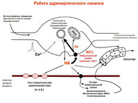
The peculiarities of the adrenergic neuron are those that can supra-linguistically long thin axons, which are formed in the organs and form dense plexuses. The total length of such axonal terminals can reach 30 cm. In the course of the terminals, there are numerical expansions - varicose veins, in which it is synthesized, stored and seen as a mediator. With the arrival of the impulse, norepinephrine is immediately seen from the numerical expansion, blowing across the great area of the smooth tongue tissue. In this way, the depolarization of m'yazovykh clitinum is accompanied by one-hour shortness of the entire organ.
Rіznі medical care, which is applied to the efector organ, similar to that of the postganglionic fiber (sympathetic, parasympathetic skinny), took away the name of mimetics (adreno-, cholinomimetics). Instructions from speech and speech, vibrating blocking the function of receptors in the postsynaptic membrane. They are called ganglion blockers. For example, ammonium vibrate vibrating H-cholinergic receptors, and atropine and scopolamine - M-cholinergic receptors.
Classical mediators win not only the function of transmitting excitement, but also the vitality and vital biological activity. The cardiovascular system is the most sensitive to acetylcholine, it stimulates and strengthens the motility of the herbal tract, simultaneously activating the activity of herbal diseases, shortening the muscles of the bronchi and decreasing bronchial secretion. Under the infusion of norepinephrine, there is an increase in the systolic and diastolic pressure without changing the cardiac rhythm, the heart contractions increase, the secretion of the tube and intestine decreases, the smooth muscles of the intestine relax. d. Adrenaline is characterized by a wider variety of ranges. For additional one-hour stimulation, ino-, chrono- dromotropic functions of adrenaline increase the heart rate. Adrenaline has a widening and anti-spasmodic action on the muscles of the bronchi, galmuic motility of the herbal tract, weakening the walls of organs, and also galmuic activity of the sphincter, secretion of the herbal tract.
Serotonin (5-hydroxytryptamine) was detected in the tissues of all animal species. In the brain, the veins are more important in the structures that regulate the regulation of visceral functions, on the periphery they are produced by enterochromaffin clitins of the intestine. Serotonin is one of the main mediators of the metasympathetic part of the autonomic nervous system, which plays a major role in neuroreflector transmission, and also functions as a mediator in the central chambers. There are three types of serotonergic receptors - D, M, T. The D-type receptors are localized mainly in the smooth meats and are blocked by lysergic acid diethylamide. The interaction of serotonin with these receptors is accompanied by mild morbidity. M-type receptors are characterized by greater autonomic ganglions; blocked by morphine. Contacting with cimi receptors, transmitting a pulsating ganglion-stimulating effect. T-type receptors, detected in the heart and leg reflexogenic zones, are blocked by thiopendol. Due to the presence of qi receptors, serotonin takes part in the development of coronary and lung chemoreflexes. Building serotonin exerts direct pressure on smooth muscles. In the vascular system, it manifests itself in constrictor and dilator reactions. With direct breathing, the muscles of the bronchi are shortened, with reflex action, the dical rhythm and legeneval ventilation change. The herbal system is especially sensitive to serotonin. On the introduction of serotonin, it reacts with a spasmodic reaction, which turns into rhythmic shortness with an increase in tone and galvanic activity, which ends.
For rich visceral organs, a characteristic purinergic transmission is named after the fact that adenosine and inosine, purine degradation products, are seen during stimulation of presynaptic terminals. The mediator in this direction is the presynaptic terminal of the effector neurons in the metasympathetic part of the autonomic nervous system.
ATP, which was seen in the synaptic gap, interacts with purinoreceptors of the postsynaptic membrane of two types. Purinoreceptors of the first type are more sensitive to adenosine, the other - to ATP. The action of the mediator is directly directed to the smooth muscles, which manifests itself in the appearance of relaxation. In the mechanism of intestinal propulsion, purinergic neurons go from the head antagonistic galvanic system to the excitatory cholinergic system. Purinergic neurons take part in the reduced low galvanization, in the mechanism of receptive relaxin, relaxation of the stravochid and anal sphincter. The shortness of the intestines, which is blamed for the purinergically crying out for relaxation, ensures the proper passage of the food breast.
Among the mediators, you can use histamine. Vіn wide extensions in various organs and tissues, especially in the herbal tract, legenia, shkіrі. Among the structures of the autonomic nervous system, the greatest amount of histamine is found in postganglionic sympathetic fibers. On the background of the reactions in certain tissues, specific histamine (H-receptors) receptors were detected: H1- and H2-receptors. Classical action of histamine and increased capillary penetration and shortness of smooth muscles. In the free state, histamine reduces the blood pressure, changes the heart rate, stimulates the sympathetic ganglion.
On the interneuronal transmission of excitation in the ganglia of the autonomic nervous system, the galvanic injection of GABA can be used. As a mediator, you can take the fate of the guilty presynaptic galvanization.
Large concentrations of various peptides, especially substance P, in the tissues of the herbal tract, hypothalamus, posterior cortex of the spinal cord, as well as the effects of stimulation of the remaining and other indications, became the basis for the use of substance P as a mediator of sensitive nerve cells.
The number of classical mediators and "candidates" in mediators, in the regulation of the activity of vicarious organs in the bere, there is a large number of biologically active speeches - mystic hormones. The stench regulates the tone, corrects the influx on the activity of the autonomic nervous system, and plays an important role in the coordination of neurohumoral transmission, in the mechanisms of vision of that diy mediators.
In the complex of active factors, prostaglandins occupy the space, which are richly located in the fibers of the bulging nerve. Sounds of stench are seen spontaneously under a burst of stimulation. The main class of prostaglandins: E, G, A, B. The main effect is the stimulation of smooth mucous membranes, suppression of the mucosal secretion, relaxation of the muscles of the bronchi. On the cardiovascular system, the stench can be differently directed: prostaglandins of class A and E call vasodilation and hypotension, class G - vasoconstriction and hypertension.
The synapse of the ANS is like a dream, like a central one. Significant expansion of chemoreceptors in postsynaptic membranes is indicated. The transmission of nerve impulses from preganglionic fibers to the neurons of all autonomic ganglia is mediated by H-cholinergic synapses, tobto. synapses on the postsynaptic membrane of such rota- tions of nicotine-sensitive cholinergic receptors. Postganglionic cholinergic fibers work on the clitins of the visceral organs (salt, SMC of the organs of etching, vessels thin) M-cholinergic synapses. It is a postsynaptic membrane that resists muscarinic receptors (atropine blocker). In these other synapses, the transmission of arousal is mediated by acetylcholine. M-cholinergic synapses may have a stimulating infusion on the smooth mucus of the herbal canal, the siechovivid system (sphincter sphincter), the veins of the SCT. However, the stinks change the vigilance, the conductivity, the shortness of the heart meat, and the crying out of the relaxation of some vessels of the head and pelvis.
Postganglionic sympathetic fibers establish 2 types of adrenergic synapses on the efectors - a-adrenergic and b-adrenergic. The postsynaptic membrane of the first a1-i a2 is adrenoreceptors. In case of NA on a1-adrenergic receptors, there is sounding of arteries and arterioles internal organs and shkіri, shortness of the mucous membranes of the uterus, sphincteris of the mucosal-intestinal tract, and at the same time relaxing other smooth mucous membranes of the herbal canal. Postsynaptic b-adrenergic receptors are also subdivided into b1 - and b2 - tips. b1-adrenergic receptors roztashovani in clitins of the heart meat. When they are affected, the alertness, conductivity and shortness of cardiomyocytes are observed. Activation of b2-adrenergic receptors leads to expansion of the vessels of the leg, heart and skeletal ulcers, relaxation of smooth bronchial ulcers, sich michur, galvanization of motility of organs etching
In addition, the postganglionic fibers were revealed, which work on the clitins of the internal organs in the histaminergic, serotonergic, purinergic (ATP) synapses.
2. Pavlov's opinion about 1 and 2 signaling systems.
The signaling system is a system of mental and insane reflex connections of the higher nervous system of creatures (including humans) and the superfluous world. Rozrіznyayut pershu and other signaling systems.
The term was approved by Academician I. P. Pavlov.
The first signaling system is practically found in all creatures, although the other system is present only in humans and, perhaps, in some whale-like ones. Tse pov'yazano z tim, scho only people zdatna shaping an abstract image of the situation. If you remember the word “lemon”, a person can show, which wine is sour and how to frown, if you hear it, then the memory of the word calls out an image in memory (the other signal system is used); yakscho with whom began the movement of the line, the robot of the first signal system.
The subject of the study of physiology is the higher nervous activity.
Another signaling system is a special type of the greater nervous activity of a person, the system of "signal signals", which goes in the direction of the wild (albeit not the same) with the creatures of the first signal system - it is evident, manifested, that it lies to the point of light. To go, like a different signal system, like a semiotic system of significances - that “what to go to the cortex of the species of our organs is other signals, signals of signals. The stench is a double-dealing act and is allowed to become aggravated, like to become our special person, especially human, more of a mind, which creates a backbone of human empirism, and, nareshti, and science - for the most important orien- t of human beings with science. I. P. Pavlov (1932).
The creature's brain only shows on the middle of the night, sounds and other rhymes follow; Apparently, they blame, to establish the first signal system of activity.
In the process of evolution of the creature's world at the stage of development of the cob development of the species Homo sapiens, there was a kind of signaling system, which ensures the active and collective adaptive habitual behavior, which created various, accepted for the group word of the signal system: P. Pavlova, becomes a "signal of signals". Opportuning of another signaling system - Viknnynennya Movy iov, signaling systems People's rhodiers, de Umovnі (Dovіlnі) Signal signals Іndivіda Nabuvyuti Sovnі, Princeyі Group Valida Ta Snowstick, rejuvenated on the signs of Movy in the direct Rosuminni Tsoeh's Words - TsE one IZ nivazyvіviyvіvіvіlіnіїї ї The life of the Homo genus, which is transmitted through movement from generation to generation.
At Vivchenni St. s. from. The backbone was more important than the accumulation of facts, which characterize the meaning of the fundamental functions of verbal signals, that buv - the expansion of the nerve mechanisms of the word. It has been established that the development of a word is developed as a result of the development of a system of mental connections (div. Intelligent reflection); its own meaning is like a quantity of calls, and its character is: a call, vibrations at the hour of the activity of a child, making the process of aggravation easier. When there are verbal signals, there are steady changes in alertness, great strength, frequency and trivality of electric discharges in the nerve cells of the singing punctures of the brain. Rosvitok St. s. from. - the result of the activity of all measles of the great pivkul; it is impossible to impair this process from the function of such an impure brain. Dosledzhennyah V.S.S. in the laboratory of higher neurodynamics and psychology of higher cognitive processes. I. Boyka showed the validity of the wedding I. P. Pavlova about dynamic timcha connections V.s.s. At the development of ideas I. P. Pavlova and E. A. Boyka at the school Y. А. the nature of the connection of the promotion with a thought, the promotion with the special promotion; SPECIALIY DISTRIBUTION OF THE DIYTYACHO MOVA TA II THERE IS ROSERBLENIY NOVIE METHODS AN ANALIZA OF PUBLICHNYY VOTTERIV (Інтент-аниназЗ), ШОО тозвольёє Стойтую мирома морова мойтовать толові товітой простовованості, ї ї dynamik, specialty of the confline situ, at Vilny hromadsky ledges and others.
Іstotnim reserve for the away doslіdzhen zalishayutsya problemi tipologії kolosalnye іndivіdualnih vіdmіnnostey in vzaєmozv'yazkah zagalnogo that spetsіalnogo tipіv GNI neocortex that emotsіyno-volovoї that mimovіlnoї regulyatsії dіyalnostі that spіlkuvannya, Pokey scho yak poorly represented in fіzіologії GNI, so i in psiholіngvіstichnih doslіdzhennyah that in antropologіchnіy lines.
Details
Ganglia be themselves cluster of multipolar (one axon and sprat dendrites) neurons(Vid kіlkoh kіtin up to tens of thousands). The extraorganic (sympathetic) ganglions may be well-displayed good tissue capsule, as a continuation of the perineurium. Parasympathetic ganglions chirp, sound, in intramural nerve gossip. Ganglia of intramural plexuses, like other vegetative knots, avenge vegetative neurons of reflex arcs. Multipolar neurons with a diameter of 20-35 µm are diffusely spread, skin neurons are drained by ganglion gliocytes.
Besides, described neuroendocrine, chemoreceptor, bipolar, and in some spinal and unipolar neurons. In sympathetic ganglia there are intensely fluorescent cells (MIF-cells) with short growths and a large number of granular bulbs near the cytoplasm. The stench sees catecholamines and chimes in on the transmission of impulses from preganglionic nerve fibers to a sympathetic efferent neuron. Cells are called interneuron.
Among the great multipolar neurons vegetative ganglion razrіznyayut: ruhovі (Clintini Dogel 1st type), sensitive (Clintini Dogel II type) and associative (Clіtini Dogel III type). Motor neurons develop short dendrites with lamellar extensions ("receptive maidans"). The axon of the clitin is arched, extends beyond the interganglion at the warehouse of thin postganglionic non-myelinated nerve fibers and terminates on smooth myocytes of the internal organs. Clitini of the 1st type are called pre-axonal neurons. Neurons of the II-th type - equal nevnovidrostkovy nerve cells. There are 2-4 growths in the upper body, and the axon is important in the middle. Not razgaluzhuyuchis, the teenagers go far in the body of the neuron. Their dendrites may be sensitive to nerve endings, and the axon will terminate on the bodies of rukhovy neurons in the vascular ganglia. Type II cells are sensitive neurons of the muscular reflex vegetative arcs. Dogel's cells of the III type are similar in shape to the vegetative neurons of the II type, these dendrites do not extend between the ganglion, and the neurite is straight up to the minor ganglia. A lot of famous people are encouraged by different types of sensitive neurons.
Thus, in the peripheral vegetative ganglia, there are macular reflex arcs, which are composed of sensitive, rough and, possibly, associative vegetative neurons.
Intramural vegetative ganglions in the walls of the herbal tract are resurrected by the same, in their warehouse, a number of rukhovy cholinergic neurons, and galvanic neurons. The stench is represented by adrenergic and purinergic nerve cells. The remaining mediator is a purine nucleotide. In the intramural vegetative ganglia, peptidergic neurons also grow, which see the vasointestinal peptide, somatostatin and a number of other peptides, for the help of which, neuroendocrine regulation and modulation of the activity of tissues and organs in the herbal system are affected.
Acetylcholine- nicotinic (curare block, hexamethonium), muscarinic (atropine block) receptors. Receptor activation → EPSP generation. Shvidky EPSP (N-cholinocer)→opening of ionic channels. Increased EPSP (M-cholinergic)→prignichennya M-strum, zumovlenogo advancement of K-conductivity.
Neuropeptides- Work like neuromodulators.

Enkefalini, speech P, luliberin, neurotensin, somatostatin - simp. ganglia (+Ach)
Catecholamines(NA, dopamine) – neurotransmitters of other cells due to intense fluorescence.
Neuropeptide Y, somatostatin - symptom. postganglionic.
Sympathetic postganglionics: NA, ATP, neuropeptide U.
α1→inosotoltriphosphate, diacylglycerol. α2→G-protein activation, ↓cAMP.
β→G-protein→AC→cAMP
Vinyatki: mediator Ach, muscarinic receptors.
Parasymp. postganglionic: Ach, VIP, NO, somatostatin, ATP, opioid peptides.
M1 (high sporidity to pirenzepine) - increased secretion of acid by clitins of the duct, M2 (low) - uplifts the heart. rhythm, secretion of mucus and sloughs.
Riznomanitna diya:
-Specific second. intermediaries: M2 may act IP3, and may in-gt AC, changing cAMP.
-Diya on Kta Sa-channels
- NO → guanylate cyclase → cGMP → cGMP-depleted protein kinase → weakened mucosal release is absorbed on the endothelium.
In the autonomic nervous system separate the central and peripheral branches. The central branches of the sympathetic nervous system are represented by the nuclei of the calf horns of the thoracolumbar branch of the spinal cord. In the parasympathetic nervous system, the central nerves include the nuclei of the middle and dovetail brains, as well as the nuclei of the biceps horns of the sacral spinal cord. Parasympathetic fibers of the craniobulbar ventricle emerge from warehouses III-ї, VII-ї, IX-ї and X-a couple cranial nerves.
Peripheral signs of the autonomic nervous system filled with nerve stovburs, ganglia and gossip.
Vegetative reflex arcs they start with a sensitive neuron, which lies near the spinal node (ganglion), like in the somatic reflex node. Associative neurons perebuvayut in the horns of the spinal cord. Here, the nerve impulses are relayed to the proximal preganglionic neurons, the adolescents of which deprive the central nucleus and reach the autonomic ganglia, where they transmit the impulses to the rough neuron. In the junction with cym, the nerve fibers of preganglionic and postganglionic are separated. The first of them deprive the central nervous system at the warehouse of the ventral roots of the spinal nerves and cranial nerves. Both in sympathetic and parasympathetic systems, preganglionic nerve fibers overlap cholinergic neurons. Axons of neurons, spread in vegetative ganglia, are called postganglionic. The stench does not make direct contact with effector cells. These terminals form expansions - varicose veins at their own pace, at the warehouse of which the bulbs of the mediator are rebuyed. In the area of varicose veins, there is no glial tunic and a neurotransmitter, seen in the middle of the esophagus, flows into the effector cells (for example, on the clitinis, smooth myocytes, etc.).
in the peripheral ganglia The sympathetic nervous system, as a rule, is known to have adrenergic efferent neurons (for a few neurons, which can synaptic connections with sweat deposits, desympathetic neurons are cholinergic). In parasympathetic ganglia, the afferent neurons are usually cholinergic.
Gangliaє accumulation of multipolar neurons (a number of cells up to tens of thousands). The extraorganic (sympathetic) ganglions may be well-displayed good tissue capsule, as a continuation of the perineurium. Parasympathetic ganglions chirp, sound, in intramural nerve gossip. Ganglia of intramural plexuses, like other vegetative knots, avenge vegetative neurons of reflex arcs. Multipolar neurons with a diameter of 20-35 µm are diffusely spread, skin neurons are drained by ganglion gliocytes. In addition, neuroendocrine, chemoreceptor, bipolar, and in some spinal and unipolar neurons have been described. In sympathetic ganglia there are intensely fluorescent cells (MIF-cells) with short growths and a large number of granular bulbs near the cytoplasm. The stench sees catecholamines and chimes in on the transmission of impulses from preganglionic nerve fibers to a sympathetic efferent neuron. Cells are called interneuron.
Among the great multipolar neurons vegetative ganglions are divided into: rukhovi (Dogel's cells of the I-th type), sensitive (Dogel's cells of the II-th type) and associative (Dogel's cells of the III-rd type). Motor neurons develop short dendrites with lamellar extensions ("receptive maidans"). The axon of the clitin is arched, extends beyond the interganglion at the warehouse of thin postganglionic non-myelinated nerve fibers and terminates on smooth myocytes of the internal organs. Clitini type I are called pre-axonal neurons. Type II neurons - rіvnovidrostkovі nerve cells. There are 2-4 growths in the upper body, and the axon is important in the middle. Not razgaluzhuyuchis, the teenagers go far in the body of the neuron. Their dendrites may be sensitive to nerve endings, and the axon will terminate on the bodies of rukhovy neurons in the vascular ganglia. Clitini type II - sensitive neurons of the muscular vegetative reflex arcs. Type III Dogel's cells are similar in body shape to type II vegetative neurons, these dendrites do not extend between the ganglion, and the neurite is straight up to the minor ganglia. A lot of famous people are encouraged by different types of sensitive neurons.
In such a manner, peripheral autonomic gangliaє mіstsevі reflex arcs, like s sensitive, rukhovih i, possibly, associative vegetative neurons.
Intramural autonomic ganglia at the station of the herbal tract, there are those, which are in the warehouse, a crim of rukhovy cholinergic neurons, є galmіvnі neurons. The stench is represented by adrenergic and purinergic nerve cells. The remaining mediator is a purine nucleotide. In the intramural vegetative ganglia, peptidergic neurons also grow, which see the vasointestinal peptide, somatostatin and a number of other peptides, for the help of which, neuroendocrine regulation and modulation of the activity of tissues and organs in the herbal system are affected.
Basic video anatomy of the autonomic nervous system (ANS)
In case of problems from a glance, download the video from the sideAvtonomnі (vegetativnі) nervovі vuzli (ganglії) mozhut roztashovuvatisya ridge vzdovzh (paravertebralnі ganglії) abo popered Demba (prevertebralnі ganglії) and takozh in stіntsі organіv: sericite, bronhіv, grass path, sechovogo mіhura that іnshih (іntramuralnі ganglії) abo nearby їh poverhnі . Sometimes stinks may look different (from decal clitin to tens of clitin) clusters of neurons, spreading along the course of some nerves or intramurally (microganglia). Preganglionic fibers (myelin) go up to the vegetative nodes to avenge the growths of cells, the bodies of which lie near the central nervous system. Qi fibers strongly flex and establish numerical synaptic termination on clitins of vegetative nodes. The reason for this is the convergence of a large number of terminals of preganglionic fibers on the skin neuron of the ganglion. In connection with the presence of synaptic transmission, the vegetative nodes are brought to the nerve centers of the nuclear type.
Vegetative nerve nodes according to the functional sign and localization are subdivided into:
pretty;
parasympathetic.
Sympathetic nerve nodes(para-prevertebral) cut off preganglionic fibers from clitin, spread in the vegetative nuclei of the thoracic and transverse segments of the spinal cord. The neurotransmitter of preganglionic fibers is acetylcholine, and of postganglionic fibers - norepinephrine (because of sweat sweat and other blood vessels, which may sympathetic cholinergic innervation). Krym tsikh neurotransmitters, enkephalins, speech P, somatostatin, cholecystokinin appear in the nodules.
Parasympathetic nerve nodes(intramural, which lie near the organs or the nodes of the head) trim the preganglionic fibers from the clitin, which are spread in the vegetative nuclei of the ovary and middle brain, as well as the cranial cord of the spinal cord. Qi fibers fill the central nervous system at warehouses 3, 7, 9, 10 pairs of cranial nerves and anterior corinths of the cranial segments of the spinal cord. The neurotransmitter of pre-postganglionic fibers is acetylcholine. The role of mediators in these ganglia is played by serotonin, ATP, and possibly peptides.
Most of the internal organs can have subvegetative vegetative innervation, so that postganglionic fibers can be removed from clitins, spread like in sympathetic and parasympathetic nodes. Reactions, mediated by clitins of sympathetic and parasympathetic nodes, often lead to prolongation, for example: sympathetic stimulation is strong, and parasympathetic - galmues heart activity.
The central plan of the existence of sympathetic and parasympathetic nerve nodes is similar. The vegetative vuzol is covered with a splenic tissue capsule and diffusely or with groups of rotting of the body of multipolar neurons, their growths in apparently unmyelinated or, more likely, myelinated fibers and endoneurium. The body of the neurons may have an irregular shape, eccentrically spread the nucleus, sharpen (sound not exactly) with shells from the glial clitin-satellites (mantle gliocytes). Often there are rich-nuclear and polyploid neurons.
Intramural junctions and connections with them, leading paths through their high autonomy, folding organization and specificity of the mediator exchange, are seen by some authors in an independent metasympathetic branch of the autonomic nervous system. Zokrema, the total number of neurons in the intramural nodes of the intestine is higher, lower near the spinal cord, and due to the folding of their interdependence in the regulation of peristalsis and secretions, they are related to the minicomputer.
Three types of neurons are described in intramural nodes:
Long-axon afferent neurons (Dogel's type I cells) are numerically superior. These are large or middle expansions of the afferent neurons with short dendrites and a long axon, which is straight behind the border to the working organ, on the clitins of which veins it establishes rukhovi or secretory endings;
Rivnovidrost part of the afferent neurons (Dogel's type II clitiny) to eliminate the long dendrites and axon, which go beyond the intervening ganglion in the suture and establish synapses on clitiny I and III types. Qi clitiny, maybe, enter like a receptor lanka to the warehouse of the mast reflex arcs, yaki close without a nerve impulse entering the central nervous system. The presence of such arcs is confirmed by the preservation of functionally active afferent, associative and afferent neurons in transplanted organs (for example, the heart);
associative clitins (Dogel's clitins of the III type) are the male insertion neurons, which engender sprats of type I and II clitins, morphologically similar to Dogel's clitins of type II. The dendrites of these clitins do not protrude beyond the internodes, but the axons straighten to other nodes, establishing synapses on type I clitins.